Services
Services We Offer
At On-Site Medical Imaging, we offer radiology (x-ray), ultrasound, vascular, cardiac, abdomen, EKG, Holter monitor, pulmonary function testing, bone densitometry, and dysphagia systems test.

Radiology (X-Ray)
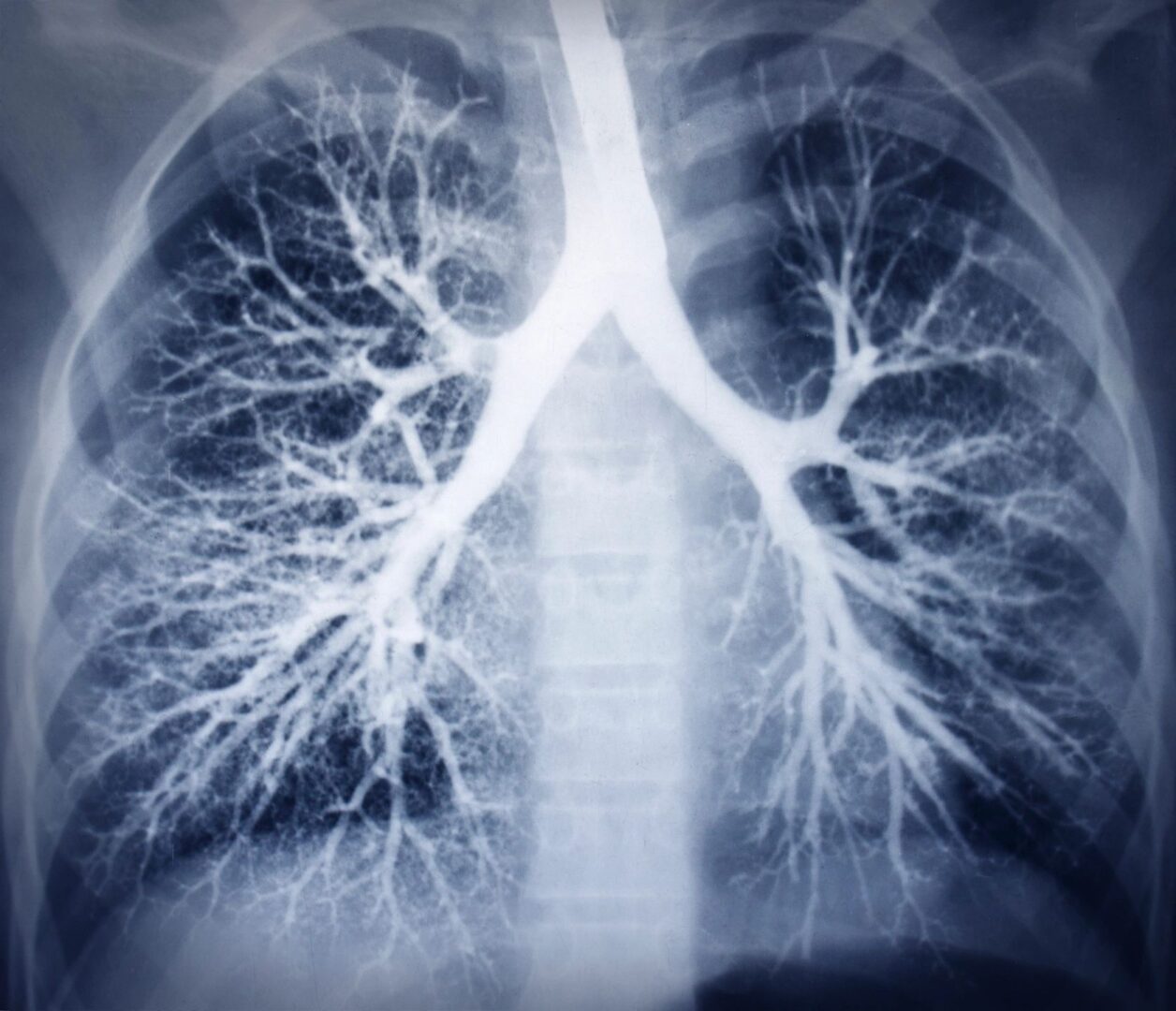
An x-ray (radiograph) is a non-invasive medical test that helps physicians diagnose and treat medical conditions. Imaging with x-rays involves exposing a part of the body to a small dose of ionizing radiation to produce pictures of the inside of the body. X-rays are the oldest and most frequently used form of medical imaging.
Ultrasound
Ultrasound imaging, also known as ultrasound scanning or sonography, is a method of obtaining images from inside the human body through the use of high-frequency sound waves. The soundwaves are recorded and displayed as a real-time visual image. No ionizing radiation is involved in ultrasound imaging.
Vascular
Vascular ultrasound provides pictures of the body’s veins and arteries. A Doppler ultrasound study may be part of a vascular ultrasound examination. Doppler ultrasound is a special ultrasound technique that evaluates blood velocity as it flows through a blood vessel, including the body’s major arteries and veins in the abdomen, arms, legs, and neck.
Cardiac
An echocardiogram is a test in which ultrasound is used to examine the heart. In addition to providing single-dimension images, known as M-mode echo that allows accurate measurement of the heart chambers, the echocardiogram also offers far more sophisticated and advanced imaging. This is known as two-dimensional (2D) Echo and is capable of displaying a cross-sectional “slice” of the beating heart, including the chambers, valves, and the major blood vessels that exit from the left and right ventricle.
Abdomen
An abdominal ultrasound produces a picture of the organs and other structures in the upper abdomen, including kidneys, liver, gallbladder, pancreas, spleen, and abdominal aorta.

EKG
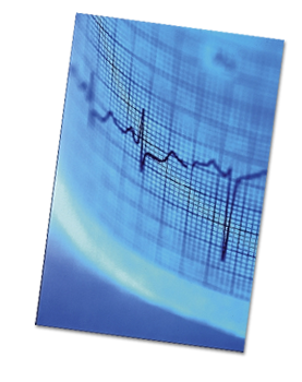
Also called Electrocardiogram or ECG, an EKG is a test that records the electrical activity of the heart that is used in diagnosing some heart abnormalities.
Holter Monitor
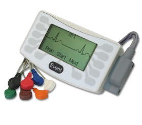
A Holter monitor is a continuous tape recording of a patient’s EKG for 24 hours. Since it can be worn during the patient’s regular daily activities, it helps the physician correlate symptoms of dizziness, palpitations (a sensation of fast or irregular heart rhythm), or blackouts.
Since the recording covers 24 hours, continuously, Holter monitoring is much more likely to detect an abnormal heart rhythm when compared to the EKG, which lasts less than a minute.
It can also help evaluate the patient’s EKG during episodes of chest pain. During this time, there may be significant changes to suggest ischemia (pronounced is-keem-ya) or reduced blood supply to the muscle of the left ventricle.

Pulmonary Function Testing
Pulmonary function tests are a group of procedures that measure the function of the lungs, revealing problems in the way a patient breathes. The tests can determine the cause of shortness of breath and may help confirm lung diseases, such as asthma, bronchitis, or emphysema.
The tests also are performed before any major lung surgery to make sure the person won’t be disabled by having a reduced lung capacity.
Bone Densitometry
Bone density scanning, also called dual-energy x-ray absorptiometry (DXA) or bone densitometry, is an enhanced form of x-ray technology that is used to measure bone loss. DXA is today’s established standard for measuring bone mineral density (BMD).

Dysphagia Systems Test
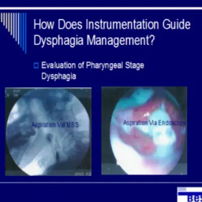
DST or Dysphagia Systems Test is a comprehensive dysphagia evaluation and therapeutic ‘all-inclusive’ session. It is performed by a clinically privileged Speech and Language Pathologist to provide information to a skilled care facility. It enables more effective management of the patient’s dysphagia–related risks.
The DST is a portable procedure that can be completed in the clinic, office, at the bedside, or wherever the patient needs to be examined. Following a thorough dysphagia evaluation process, a fiberoptic endoscope is passed trans nasally, permitting inspection of swallowing mechanisms and functions from the velopharynx to hypopharynx and larynx.
Status of standing secretions in the hypopharynx, frequency effectiveness of spontaneous swallowing, and potential implications for aspiration is taken into consideration as the exam proceeds.
The patient is then directed to perform various tasks to evaluate the sensory and motor status of the pharyngeal and laryngeal mechanism. Laryngeal competence is assessed by observing volitional, maintained closure of the airway as a potential airway protection mechanism.